
Classification of rhinoplasty according to the thickness of soft tissues
Preoperative classificatory system that differentiates two types of patients according to the thickness of the nasal skin envelope and guides surgical planning and the indication of regenerative adjuvants.
Why we measure the thickness of your skin before surgery
The classification is based on a simple fact – the thickness of the nasal skin – but has important implications for the final outcome of the surgery.
– 01 / For the patient
What is this classification and why does it matter?
Before scheduling a rhinoplasty, at Mallorca Medical Group we measure the thickness of the skin of your nose with a simple instrument called an adipometer. This measurement, which is obtained in seconds and painlessly during the first consultation, is one of the most useful pieces of information to predict how your nose will behave after surgery.
A rhinoplasty, to simplify, modifies the bony and cartilaginous structure that shapes the nose. But that structure is covered by a skin that acts as a veil: the thinner the skin, the more transparent what is underneath; the thicker the skin, the more it dampens the underlying relief and the more it tends to swell over a long period of time.
It is not a question of “good skin” or “bad skin”. Both skins have their own predictable behaviors, each with its advantages and challenges. What changes is how we plan the surgery so that the result is optimal in each case.
Why is this data so important?A technically flawless rhinoplasty can give a disappointing result if we have not correctly anticipated how the skin will respond. Grading allows us to anticipate that response and plan the surgery accordingly, rather than finding out months later.
– 02 / For the patient
The two types: thin skin and thick skin
Measuring the thickness of the nasal skin envelope allows us to classify the patient into one of two types. The threshold between the two is 3 millimeters measured at the rhinion (the bony point of the nasal dorsum).
Typical behavior
The skin reveals the relief of the underlying skeleton. Any slight irregularity of the bone or cartilage tends to become visible after a few months, once the postoperative period has subsided.
Principal risk
- Appearance of visible irregularities on the dorsum of the nose
- Visualization of bony rims or subtle asymmetries
- Late dissatisfaction despite good immediate surgical outcome
Recommended strategy
Duvet Rhinoplasty: intraoperative application of a biologic nanogreasy layer enriched with L-PRF that acts as a regenerative padding and prevents the transparency of irregularities.
Typical behavior
The skin cushions the underlying relief and masks irregularities well, but tends to a more prolonged inflammatory response and fibrous scarring that can detract from the definition of the nasal contour.
Principal risk
- Prolonged postoperative inflammation
- Loss of contour and tip definition
- Final result less refined than desired
Recommended strategy
Surgical maneuvers oriented to the definition of the nasal contour and tip, together with postoperative follow-up adapted to accompany the natural evolution of the healing process.
– 03 / For the patient
How is it measured?
The measurement is simple, quick and painless. It is performed during the first consultation with an instrument called a manual adipometer, similar to the one used in nutritional consultations to measure skin folds.
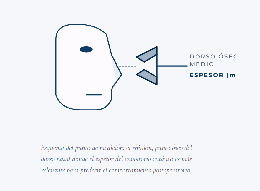
The measurement procedure is as simple as that:
- The doctor applies the adipometer to the skin of the rhinion (the bony spot on the back of your nose), without pressing uncomfortably.
- The instrument measures skinfold thickness in millimeters.
- The values are noted and classified as Type I (< 3 mm) or Type II (≥ 3 mm).
- This information, together with the rest of the assessment, defines the personalized surgical plan.
The measurement does not require any prior preparation on your part and is done in the same session as the first consultation. If in your case the result is just at the threshold (around 3 mm), we will evaluate other factors – history, general characteristics of the facial skin, aesthetic objective – to decide which of the two strategies is best suited.
– 04 / For the patient
What does it mean for my treatment?
The classification does not change the type of surgery performed – it will still be a rhinoplasty, with its corresponding surgical steps – but the complementary decisions that are made around it:
- If it is Type I (thin skin), planning will include Duvet Rhinoplasty with biologic nanogreasy and L-PRF mantle, to prevent visible dorsal irregularities. The surgery is likely to be about 30-45 minutes longer because of the preparation of adjuvant materials.
- If it is Type II (thick skin), the planning will prioritize the technical maneuvers to maximize the definition of the nasal contour and tip. The postoperative period may require longer follow-up to follow the natural evolution of healing, which is characteristically slower in this type of wrap.
In both cases the objective is the same: to obtain the best possible result adapted to your anatomy, not to apply a single technique that fits all patients equally.
Technical-scientific block
Rationale, classification criteria, measurement methodology, therapeutic implications and limitations of the classification system.
– 05 / Technical block
Basis of the classification system
The postoperative behavior of the nasal skin envelope is one of the factors with the greatest impact on the final aesthetic outcome of rhinoplasty, yet it has historically received less attention than bone and cartilage remodeling. Modern surgery has refined protocols for nasal skeletal modification, but the criteria for anticipating and modulating the envelope response have traditionally been more subjective.
The proposed classification is based on a reproducible clinical observation: the thickness of the soft tissue of the nasal dorsum is the most consistent predictor of postoperative behavior, both in terms of the transparency of the underlying relief and the inflammatory and scar response. This observation, contrasted in our own clinical series, motivated the development of a dichotomous system that makes it possible to convert a qualitative assessment into a measurable and reproducible variable.
The threshold of 3 mm was established from the analysis of the clinical series of Dr. Garcia Ceballos as a turning point in the behavior of the envelope: below this value, the risk of transparency of irregularities predominates; above this value, the risk of prolonged inflammation and fibrous scarring predominates.
– 06 / Technical block
Measurement methodology
Instruments
Standard manual adipometer, similar to the one used in nutritional anthropometry for skinfold measurements. The choice of instrumentation responds to criteria of simplicity, reproducibility and universal availability in the office, without the need for specific technology.
Anatomical reference point
Rhinion, the middle bony point of the nasal dorsum corresponding to the junction of the nasal bone with the upper lateral cartilages. This location has been selected because:
- This is the area of maximum visual exposure of the nasal dorsum and, therefore, the most relevant for the perceived esthetic result.
- It presents less anatomical variability between patients than the more distal areas (supratip, tip).
- This is the region where the main bone remodeling is performed and where the biological mantle is applied in Type I patients.
Measurement procedure
- Patient in seated position, head in Frankfurt plane.
- Palpatory identification of the rhinion.
- Gentle pinching of the skin fold with the adipometer, without excessive compression of the tissue.
- Direct reading in millimeters after stabilization of the value (typically 2-3 seconds).
- Repeat measurement and averaging of values to reduce variability.
– 07 / Technical block
Classification and classification criteria
| Type | Thickness | Expected behavior | Surgical strategy |
|---|---|---|---|
| Type I | < 3 mm | Increased risk of transparency of irregularities of the bony and cartilaginous skeleton. Generally favorable healing. Rapidly appreciable postoperative result. | Rhinoplasty + Duvet Rhinoplasty (biological nanogreasy + L-PRF). Emphasis on careful bone polishing and smoothness of the reconstructed contour. |
| Type II | ≥ 3 mm | Lower risk of transparency of irregularities. Tendency to prolonged inflammation and subcutaneous fibrosis. Potential loss of contour and tip definition. | Rhinoplasty with emphasis on contour and tip definition maneuvers. Longer postoperative follow-up, adapted to the characteristic inflammatory behavior of the thick envelope. |
Borderline cases and patients at the threshold
In patients with values close to 3 mm, the decision is individualized considering additional factors:
- History: previous rhinoplasty, trauma, previous skin infections that may have modified the envelope.
- General characteristics of facial skin: seborrhea, predisposition to fibrosis, scar quality documented in other interventions.
- Aesthetic objective: magnitude of the desired change, need for extensive bone modifications.
- Age and skin elasticity: a modulating factor in the behavior of the envelope.
– 08 / Technical block
Therapeutic implications
In Type I patients
The main indication is the intraoperative application of the biological mantle of Duvet Rhinoplasty (L-PRF-enriched nanofat), applied on the remodeled nasal dorsum before skin closure. The objective is twofold: mechanical prevention of the transparency of irregularities in the short and medium term, and regenerative improvement of the skin envelope in the long term.
Additionally, in this group, priority is given to meticulous bone polishing, avoiding leaving abrupt edges or transitions in the underlying skeleton that could become transparent despite the biological covering.
In Type II patients
The strategy focuses on surgical maneuvers aimed at maximizing contour definition and postoperative management adapted to the expected behavior of the envelope:
- Surgical maneuvers to enhance tip definition and dorso-supratip transition.
- Specific attention to the prolonged inflammatory behavior characteristic of this type of envelope.
- Longer postoperative follow-up, with staggered visits during the first year, to follow the natural evolution of the healing process.
Compatibility with basic rhinoplasty techniques
The classification is independent of the surgical approach used (open vs. closed), the underlying technique (structural, sparing, conservative) and the instrumentation used (conventional rhinoplasty, ultrasonic). It functions as an additional layer of planning that is superimposed on the usual technical decisions.
– 09 / Technical block
Recognized limitations and future development
- Simplified dichotomous system. The classification into two types prioritizes clinical utility and reproducibility over granularity.
- Single variable considered. The thickness of the envelope is a relevant but not the only predictor. Other variables – cutaneous elasticity, vascularization, periosteal status – may modulate postoperative behavior.
- Operator-residual measurement dependence. Although the adipometer reduces subjectivity, the measurement retains an operator-dependent component. Standardization would require specific training protocols.
- Evidence of a single surgeon. The system was developed and validated in the author’s own clinical series. Its external validation by independent teams would be desirable.
- Absence of comparison with advanced instrumental techniques. High-frequency cutaneous ultrasound would allow more accurate thickness measurements. Cross-validation between adipometer and instrumental techniques would be useful.
Despite these limitations, the classification fulfills its main objective: to offer the surgeon a simple, reproducible tool, applicable in the office without additional equipment, which improves surgical planning with respect to subjective qualitative assessment.
— 10
Associated resources
Frequently Asked Questions
The thickness of the nasal skin determines how the dorsum behaves after rhinoplasty. Thin skins are at greater risk of revealing irregularities of the underlying bony or cartilaginous skeleton; thick skins are more prone to fibrous scarring and prolonged inflammation. Classifying the patient before surgery makes it possible to anticipate these behaviors and to plan the surgical technique and regenerative adjuvants in a personalized way.
By means of a manual adipometer, a simple instrument similar to the one used in nutrition to measure skin folds. The measurement is performed in the office, is painless and takes seconds. It is measured in the middle bone dorsum, where skin thickness is more relevant to predict postoperative behavior.
Type I corresponds to patients with thin skin (thickness less than 3 mm). In this group the risk of visible dorsal irregularities after surgery is high. Type II corresponds to patients with thick skin (3 mm or more); in this group the risk of irregularities is lower but there is a greater tendency to prolonged inflammation and loss of definition. Each type receives an adapted treatment.
It does not change the fundamental surgical procedure, which continues to be a rhinoplasty. It changes the complementary decisions: in Type I patients it typically incorporates Duvet Rhinoplasty with nanogreasy and L-PRF biological mantle; in Type II patients it prioritizes techniques to control inflammation and improve contour definition.
It is a proprietary classification developed by Dr. García Ceballos and published in 2024 in Cirugía Plástica Ibero-Latinoamericana, a peer-reviewed scientific journal with Ibero-Latin American circulation. As with any recent scientific contribution, its international adoption will depend on its reproduction and validation by other teams in the coming years.
In borderline cases the decision is individualized by evaluating other factors: history (previous rhinoplasty, trauma), aesthetic expectations, general facial skin characteristics, factors predisposing to fibrosis or atrophy. The classification is an orientative tool, not an automatism: each patient receives a personalized evaluation.
Complementary contributions to the classification
The classification is performed at the first consultation by means of a simple measurement. It will help you understand what to expect from surgery and what surgical strategy is most appropriate in your case.